
Project
Human Tenocyte metabolism under pathological and physiological loading conditions
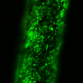
Fig 1: PEG-Peptide rod seeded with bovine tendon cells and stained with Calcein AM.
| Primary Investigator: | Dr Dharmesh Patel |
| Co-investigators: | Hazel Screen |
| Graham Riley | |
| Stephanie Bryant | |
| Funder: | Arthritis Research UK Funded: NE/PhD/19598 National Institutes of Health USA Funded: R21 |
The aim of this project is to develop a system to explore the effect of different tension and shear environments on the metabolism of human tenocytes, exploring both physiological equivalent and pathological conditions.
The Fibre Composite System
A fibre composite material to mimic the aforementioned environment has been synthesised using Poly(ethylene glycol) (PEG) as the matrix material and a peptide conjugated PEG for fibres/rods within.
Analysis of composites has shown strain exhibited by rods can be controlled in relation to gross applied strain by altering the stiffness of rods or 'soak' time during fabrication. Tenocytes are seen to attach sufficiently to rods, thus being incorporated into the composite while also remaining viable when left in PEG solution (prior to polymerisation) for up to 120 minutes.
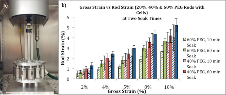
(a) Bose system with multi-chamber older. (b) Micomechanics data summary of fibre composite system. Higher PEG % result in stiffer rods. Graph shows changing either soak time or rod stiffness changes the extend of rod steain at a given applied gross strain.
The hierarchical structure of tendons leads to a complex environment for the cells within (tenocytes), consisting of both tension and shear. These are important cues in mechanotransduction where they can influence the cellular response and currently poorly understood for tenocytes.
Gene Expression Analysis
Gene expression analysis, focusing on genes potentially involved in matrix turnover, has been conducted using the different tension and shear ratios created by the composite system. Data comparing healthy and tendinopathic human tenocytes indicates that tendinopathic cells are more mechano-sensitive than healthy cells, showing larger gene expression changes in response to loading. Differences in the basal gene expression between tendinopathic and healthy tenocytes were also identified. Shear higher and lower than the physiological equivalent was found to evoke responses different from one another; healthy tenocytes downregulated matrix synthesis with high shear and upregulated matrix degradation with low shear, while tendinopathic tenocytes downregulated matrix degradation with high shear
Altering the peptide to one that mimics collagen was also found to increase the number shear-tension ratio regulated genes, suggesting integrins are involved in sensing this mechanotransduction cue.